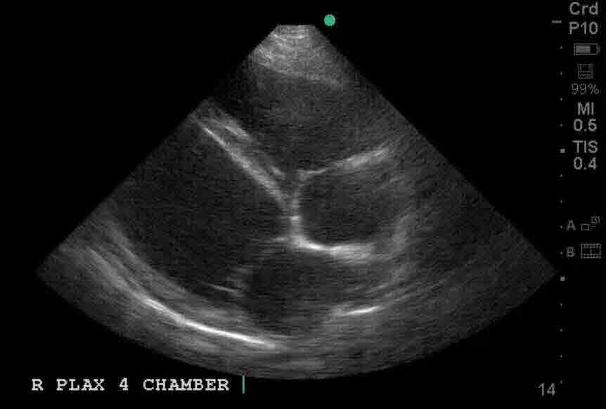
This is a great depiction of Trypanosomiasis (Chaga's Disease). Note the thin walls and dramatically reduced motion. Click here for a normal heart.
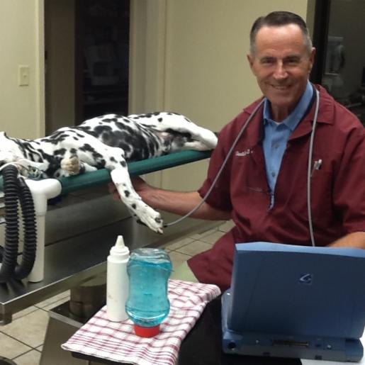
I am Dr. Charlie Wiley.
We are Alamosa Enterprises, Inc. in Alice, Texas and we offer mobile veterinary ultrasound in South Texas.
I graduated from Texas A&M College of Veterinary Medicine in 1976.
In 2010, I discovered ultrasound and dedicated my veterinary career to
that one goal. With the help of
Dr. Andra Voges and the late
Dr. Sherm Mathey along with the
radiology staff at Lackland AFB, I became skilled in ultrasound.
In 2011, I met and became a student of
Dr. Michelle Fabiani
chief radiologist at Gulf Coast Veterinary Radiology in Houston and over
the next several years, excelled under her direction which included
intense specialized training that allowed me to become a part-time
sonographer for Gulf Coast.
For the past 8 years, I have been working very closely with
Dr. Victoria McEwen,
chief radiologist at Lighthouse Radiology in Florida. Dr. McEwen reads all my abdominal scans and radiographs.
Most of my early echocardiography training was with
Dr. Adam Kane a
boarded cardiologist who was with Gulf Coast Veterinary Telemed at the time. Adam was responsible for training me to become
very proficient with comprehensive echoes, including measurements, color doppler and spectral doppler using continuous wave
and pulse wave.
Currently, I am sending my echoes to Coastal Veterinary Cardiology where
Dr. Maggie Lamy is the chief cardiologist. Maggie is also a wonderful tutor and gives me frequent instruction on how to
continue to provide the most diagnostic images.
My training experience started about 5 years ago when I was invited to be an instructor in one of Dr. Greg Lisciandro's FAST
Classes at SWVS. Since then, I had the priviledge of training for Dr. Lisciandro at SWVS for the next 3 years.
Samsung has also
hired me to provide training for new owners that have just purchased a new machine.
As of September 2026, I have done over 8,000 diagnostic ultrasounds.
My first machine was a Sonosite MicroMaxx, then an M-Turbo followed by an Edge II.
Taking advantage of new technology, I just purchased a new
Samsung HM70 EVO
system.
Why did I move to Samsung?
The new technology in the HM70 will give me additional power to do larger dogs; something that was not available with my previous
system.
If your clinic is within 3 hours of Alice, I can travel to your office! I normally cover the area south of Victoria all the way to
Brownsville.
If you are interested in either a diagnostic scan or training for your clinic, please
Click here to fill out a contact form and please include your clinic name....
Click here to submit a request for a diagnostic ultrasound.
I would love to hear from you!
Thanks and God Bless!
Case Images
Click on the image to enlarge it
For videos, set the play to "Loop" for a longer viewing
This is a great depiction of Trypanosomiasis (Chaga's Disease). Note the thin walls and dramatically reduced motion. Click here for a normal heart.
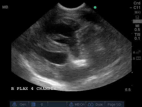
This is one of the worst cases of HW infestations that I have encountered. Note how the worms are clumped in the annulus of the tricuspid valve and the severe dilation of the RA and RV.
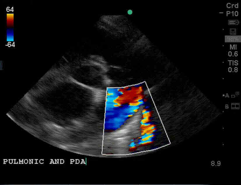
This is a very good PDA case. The view is the R PSAX HI view and the turbulence is from the patent PDA injecting directly into the MPA. The spectral doppler waveform of this abnormal flow is here, clearly demonstrating the regurgitant flow back through the pulmonic valve.
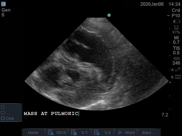
This case is a cardiac mass on the dorsal border of the pulmonic valve, causing significant obstruction to the outflow of the RVOT and a severe TR, seen here. The spectral doppler taken at the pulmonic valve, shows the distinctive "type iii" waveform here, clearly demonstrating the "notch" seen in severe PHT.
Click
here to submit
information for a new case.
May God Bless, and I hope to hear from you.
Charlie Wiley